免费一级a一片在线播放无_91精品国产91热久久久久福利_国产伦精品免编号公布_国产精品黄网站免费进入_日本韩国三级观看_久久人人妻人人爽人人卡片av_亚洲成A人无码_国产精品自产拍在线观看

所在位置: 首頁 > 公司新聞

什么是HORIBA堀場拉曼成像和光譜儀!?


 采集拉曼光譜成像和剖面圖


顯微拉曼系統(tǒng)配備XY(Z) 自動樣品平臺就能夠記錄拉曼光譜成像或剖面圖。拉曼光譜成像是一種強(qiáng)有力的技術(shù),它基于樣品的拉曼光譜得到詳細(xì)的化學(xué)信息分布圖像。在圖像的每一個像元上,都對應(yīng)采集了一條完整的拉曼光譜,將這些光譜特征集成在一起,就產(chǎn)生了一幅反映材料的成分和結(jié)構(gòu)的偽彩圖像:

  • 根據(jù)拉曼峰強(qiáng)生成材料濃度和分布圖像
  • 根據(jù)拉曼峰位生成材料的分子結(jié)構(gòu)、相以及材料的應(yīng)力變化圖像
  • 根據(jù)拉曼峰寬生成材料的結(jié)晶度和相的分布圖像


因此,根據(jù)一套數(shù)據(jù)可以構(gòu)建出多種不同的拉曼光譜成像,能給研究人員提供遠(yuǎn)超過人眼所能看到的內(nèi)容。

拉曼剖面圖和圖像可以在在一維、二維和三維方向上收集,包括:

  • 一維:線掃描、深度掃描、時間掃描、溫度掃描等
  • 二維:XY 成像、XZ 和YZ 成像
  • 三維:XYZ 數(shù)據(jù)立方

分析樣品表面以下的特征

Raman depth profile of a layered polymer structure, showing the distribution of three chemically distinct layers from the surface (left hand side) down to 40 μm below the surface (right hand side).

Raman depth profile of a layered polymer structure, showing the distribution of three chemically distinct layers from the surface (left hand side) down to 40 μm below the surface (right hand side).

只要樣品對激光是透明的,共焦顯微拉曼光譜儀就能夠用來分析樣品表面以下的特征。例如對液體 / 氣 體包裹體的分析,對玻璃內(nèi)污染物的分析以及多層聚合物材料結(jié)構(gòu)的分析。

通常在拉曼系統(tǒng)中,需要人工將激光聚焦在樣品內(nèi)某個預(yù)定位置,然后進(jìn)行光譜分析。如果顯微拉曼光譜儀配備了Z方向的自動移動控制,就可以在Z方向上自動運(yùn)動,獲得不同深度的光譜,從而得到深度方向的剖面分析。

75 μm multi-layered polymer of polyethylene-Nylon- polyethylene

75 μm multi-layered polymer of polyethylene-Nylon- polyethylene

在剖面上每一點(diǎn)對應(yīng)采集了一條完整的拉曼光譜,這些光譜集成在一起,就產(chǎn)生了一幅反映材料成分和結(jié)構(gòu)變化的曲線:

  • 根據(jù)拉曼峰強(qiáng)生成材料濃度和分布的曲線
  • 根據(jù)拉曼峰位生成材料的分子結(jié)構(gòu)、相以及材料的應(yīng)力的曲線
  • 根據(jù)拉曼峰寬生成材料的結(jié)晶度和相的曲線

額外配備 XY 自動控制樣品平臺的拉曼系統(tǒng)可以用來 在樣品中進(jìn)行光學(xué)切片,比如產(chǎn)生XZ或YZ拉曼光譜成像。

分析液體樣品

顯微拉曼光譜儀可以用來對液體樣品進(jìn)行分析,就如同分析固體、漿狀物、膠體、氣體和粉末樣品一樣。

對于非揮發(fā)性液體,可以在載玻片上滴一小滴,然后采用常規(guī)方式將激光聚焦后進(jìn)行分析。

揮發(fā)性液體( 非揮發(fā)性液體亦可) 可以裝在玻璃比色皿中,使用液體樣品池來進(jìn)行分析,它有一個多通路的反射鏡可以提高信號強(qiáng)度。另一種選擇是將液體裝在小玻璃瓶里,然后利用共焦顯微拉曼光譜儀使玻璃容器的拉曼信號干擾*小化,從而能得到容器內(nèi)液體的高質(zhì)量拉曼光譜。

拉曼光譜成像能提供的信息

Raman spectral imaging (or mapping) is a method for generating detailed chemical images based on a sample’s Raman spectrum. Raman spectral imaging allows chemical distribution to be viewed which is invisible by standard optical microscopy.

Raman spectral imaging (or mapping) is a method for generating detailed chemical images based on a sample’s Raman spectrum. Raman spectral imaging allows chemical distribution to be viewed which is invisible by standard optical microscopy.

拉曼光譜成像是一種強(qiáng)有力的技術(shù),它基于樣品的拉曼光譜生成詳細(xì)的化學(xué)圖像,在圖像的每一個像元上,都對應(yīng)采集了一條完整的拉曼光譜,然后把這些光譜集成在一起,就產(chǎn)生了一幅反映材料的成分和結(jié)構(gòu)的偽彩圖像:

  • 根據(jù)拉曼峰強(qiáng)生成材料濃度和分布圖像
  • 根據(jù)拉曼峰位生成材料的分子結(jié)構(gòu)、相以及材料的應(yīng)力圖像
  • 根據(jù)拉曼峰寬生成材料的結(jié)晶度和相的圖像

一般在拉曼光譜成像實(shí)驗中,樣品移動和光譜采集都是連續(xù)進(jìn)行的。根據(jù)用戶定義的區(qū)域范圍重復(fù)數(shù)百、數(shù)千甚至數(shù)百萬次采集數(shù)據(jù)。

拉曼光譜成像可以在二維或三維進(jìn)行收集,生成XY、XZ 或YZ 圖像,以及XYZ 數(shù)據(jù)立方。

拉曼光譜成像對于很多不同領(lǐng)域的科學(xué)家來說,都是非常有價值的技術(shù),因為它能夠顯示出普通的光學(xué)顯微鏡下觀察不到的化學(xué)成分分布。

拉曼光譜成像能提供什么樣的信息?

A Raman spectral image contains a full Raman spectrum at each and every pixel of the image. Raman spectral images provide chemical and structural information about a sample which cannot be observed using tradition light microscopy.

A Raman spectral image contains a full Raman spectrum at each and every pixel of the image. Raman spectral images provide chemical and structural information about a sample which cannot be observed using tradition light microscopy.

拉曼光譜成像上的每一個像元,都對應(yīng)于一條完整的 拉曼光譜,這些數(shù)百、數(shù)千甚至數(shù)百萬條光譜綜合在一起, 就產(chǎn)生了一幅反映材料的成分和結(jié)構(gòu)的偽彩圖像:

  • 根據(jù)拉曼峰強(qiáng)生成材料濃度和分布圖像
  • 根據(jù)拉曼峰位生成材料的分子結(jié)構(gòu)、相以及材料的應(yīng)力圖像
  • 根據(jù)拉曼峰寬生成材料的結(jié)晶度和相的圖像
Raman spectral images provide chemical and structural information about a sample which cannot be observed using tradition light microscopy.

Raman spectral images provide chemical and structural information about a sample which cannot be observed using tradition light microscopy.

拉曼光譜成像可提供樣品的化學(xué)信息和結(jié)構(gòu)信息,這 些信息是無法用傳統(tǒng)的光學(xué)顯微鏡獲得的。一般情況下,它可以用來得出:

  • 組分的分布,顆粒大小
  • 樣品中結(jié)晶度的改變、相變
  • 污染物顆粒大小和形狀
  • 不同相的邊界組分的相互作用和混合
  • 樣品的應(yīng)力分布

獲取一幅拉曼光譜成像需要多長時間?

The development of ultra-fast Raman spectral imaging modules allows drastically reduced measurement times with acquisition times down to < 1ms/point.

The development of ultra-fast Raman spectral imaging modules allows drastically reduced measurement times with acquisition times down to < 1ms/point.

拉曼光譜成像的采集時間由很多參數(shù)決定,包括成像區(qū)域大小、所需像元 ( 數(shù)據(jù)點(diǎn) ) 數(shù)目以及每個像元的采集時間 ( 取決于樣品成分的拉曼強(qiáng)度和對光譜質(zhì)量的要求 )。一般拉曼光譜成像可包含數(shù)百、數(shù)千甚至數(shù)百萬條拉曼光譜,所以采集時間可能會比較長。

采集拉曼光譜成像所需要的時間長短不一,從幾秒鐘到幾天都是有可能的,具體取決于上述各項條件。

The acquisition time for a Raman spectral image depends on many parameters, including the size of the image area, the number of pixels (data points) required, and the acquisition time per pixel.

The acquisition time for a Raman spectral image depends on many parameters, including the size of the image area, the number of pixels (data points) required, and the acquisition time per pixel.

傳統(tǒng)的拉曼光譜成像常常需要相對較長的采集時間, 但對于拉曼散射效率特別低的材料也能給出很高的靈敏度,還能提供很高的光譜分辨率,能夠測量的光譜范圍也比較 大。采用這種成像方式,一般每個數(shù)據(jù)點(diǎn)的采集時間在1 秒 ~10 秒的量級或者更高,一般總測量時間從幾小時到幾天不等。

超快拉曼光譜成像模塊的開發(fā)極大地縮短了測量時 間,其單個數(shù)據(jù)點(diǎn)的采集時間可以降低到 5 毫秒以下,這樣的速度意味著大面積細(xì)致的拉曼光譜成像也能夠在幾秒到幾分鐘內(nèi)完成。

什么是超快速拉曼光譜成像?

Ultra-fast Raman spectral imaging is a technique for allowing Raman spectral images to be acquired with acquisition times down to less than 5ms/point, resulting in total measurement times of seconds or minutes, even for images comprising tens or hundreds of thousands of spectra.

Ultra-fast Raman spectral imaging is a technique for allowing Raman spectral images to be acquired with acquisition times down to less than 5ms/point, resulting in total measurement times of seconds or minutes, even for images comprising tens or hundreds of thousands of spectra.

超快拉曼光譜成像是一種新型光譜成像技術(shù),能夠使采集拉曼光譜成像時每個數(shù)據(jù)點(diǎn)的采集時間不超過5毫秒,從而即使是包含幾萬到幾十萬條光譜的圖像,總的測量時間也只有幾秒或幾分鐘。

傳統(tǒng)的拉曼光譜成像一般需要相對較長的采集時間, 但對于拉曼散射效率特別低的材料也能給出很高的靈敏度,還能提供很高的光譜分辨率,能夠測量的光譜范圍也比較大。這種成像方式一般每個數(shù)據(jù)點(diǎn)的采集時間在 1 秒 ~10 秒的量級或者更高,從而總測量時間從幾小時到幾天不等。

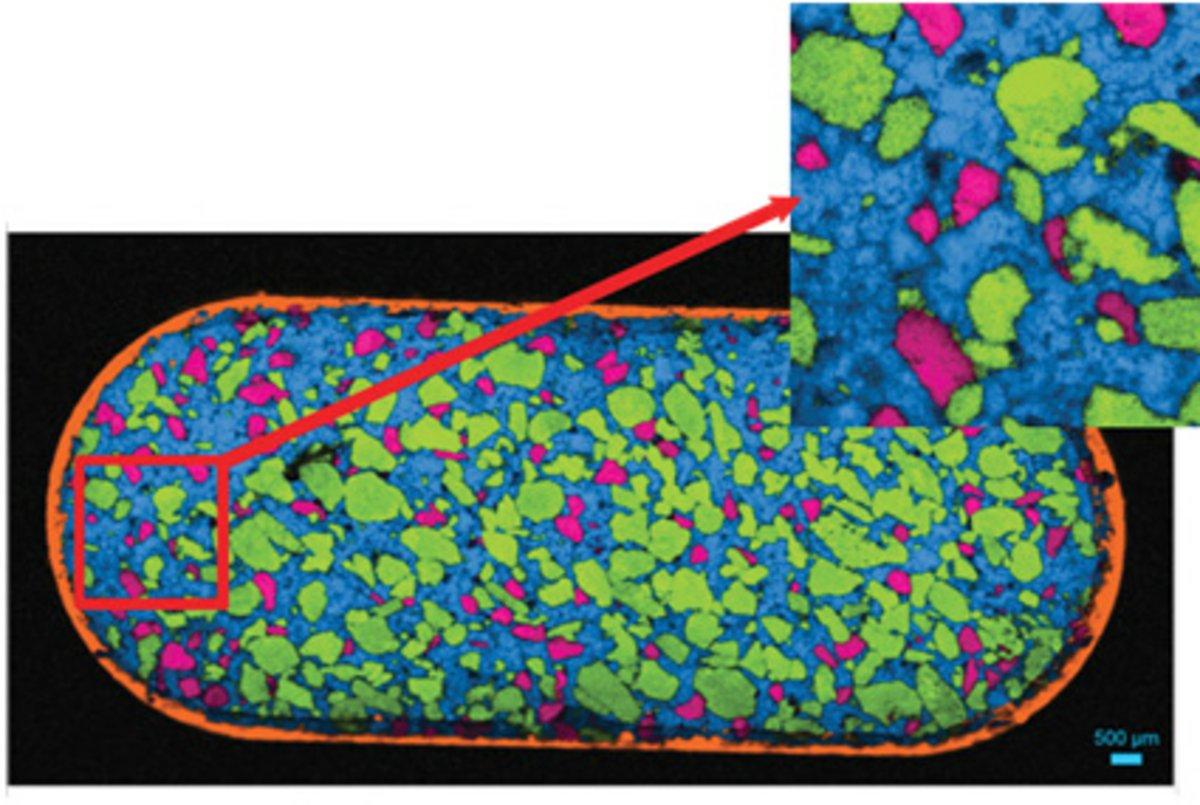
Raman spectral image of a 0.6 x 2.4 mm2 area of a pharmaceutical tablet, showing the distribution of aspirin (red), paracetamol (green),caffeine (blue) and cellulose (yellow).

Raman spectral image of a 0.6 x 2.4 mm2 area of a pharmaceutical tablet, showing the distribution of aspirin (red), paracetamol (green),caffeine (blue) and cellulose (yellow).                                                                                                            

盡管超快拉曼光譜成像的方法有很多種,但是一般都是把樣品平臺的移動以及探測器讀出相結(jié)合,使得在標(biāo)準(zhǔn)的“逐點(diǎn)”成像實(shí)驗中的“死時間”*小化。

超快拉曼光譜成像時,單個數(shù)據(jù)點(diǎn)的采集時間可以降低到 5 毫秒或者更少,這樣的速度意味著大面積精細(xì)的拉曼光譜成像也能夠在幾秒到幾分鐘內(nèi)完成。

超快拉曼光譜成像并不適合所有的樣品,其效力依賴于樣品自身的拉曼強(qiáng)度,也取決于創(chuàng)建圖像所需要的光譜質(zhì)量。                                                                                                                                                                                                                                                     

LabRAM Soleil? 高分辨超靈敏智能拉曼成像儀

LabRAM Odyssey 高速高分辨顯微共焦拉曼光譜儀

LabRAM Odyssey Semiconductor

XploRA? PLUS共焦拉曼光譜儀